 |

Neurological
Basis of Behavior (PSY -
610)
VU
Lesson26
Basic
Neuroanatomy
Objectives:
The
student would learn about the ionic and molecular movement of the neurons and
how the
electrophysiological
properties of neurons change
·
Systems, structure,
Cells of the NS Neurons, Types of neurons, axonic and
dendritic
communications,
·
Neuronal conduction
and functioning, ionic and electrophysiological properties,
·
Localizing brain
areas planes of reference (anterior-posterior etc).
·
The
Brain and the Peripheral systems: Brain: Forebrain, Mid brain, Hind Brain
functioning of
each
anatomical location in the CNS.
We
have studied in our earlier lesson how the neuronal membrane is structured, and
how the
phospholipids
form a tight mesh from which substances and molecules have difficulty leaving
or
entering.
We would discuss this more in detail
Control
of molecules:
In
the Phospholipid layers, the movement of lipid molecule through the membrane is
easier and also that
of
smaller molecules. The cell membrane also allows materials to move in and out
depending on the
changes
in the membrane permeability. Increased Permeability mean that membrane can
allow those
materials
to pass which had earlier not been able to pass through, and decreased
permeability means that
the
gates of passing in/out are closed
Membrane
permeability is determined by ionic state of membrane:
The
most important task of the neurons is to communicate, and we have seen that
neurons are active as
living
systems. There is a constant movement of ions in the intracellular and the
extracellular
membrane.
This constant state of flux in which these ions (Ions are molecules which are
negatively
charged,
or positively charged depending on the number of electrons they carry) are
moving generates
electrical
charges which then enable neurons to communicate and to send out electrical
signals.
Electrical
charges are measured in terms of
volts (milli volts in the case of neurons) and the
difference
of
electrical charge between the intracellular membrane and the extracellular
membrane is known as the
Potential
Using
a voltmeter by which we can place one electrode on the intracellular and one on
the extracellular
membrane
we would find that the inside has a large concentration of negatively charged
ions whereas
the
extracellular membrane has more positively charged ions. Thus, the inside of the
cell is negative as
compared
to outside and the difference in potential is recorded at -70 mV (this is about
1/15th of
the
difference
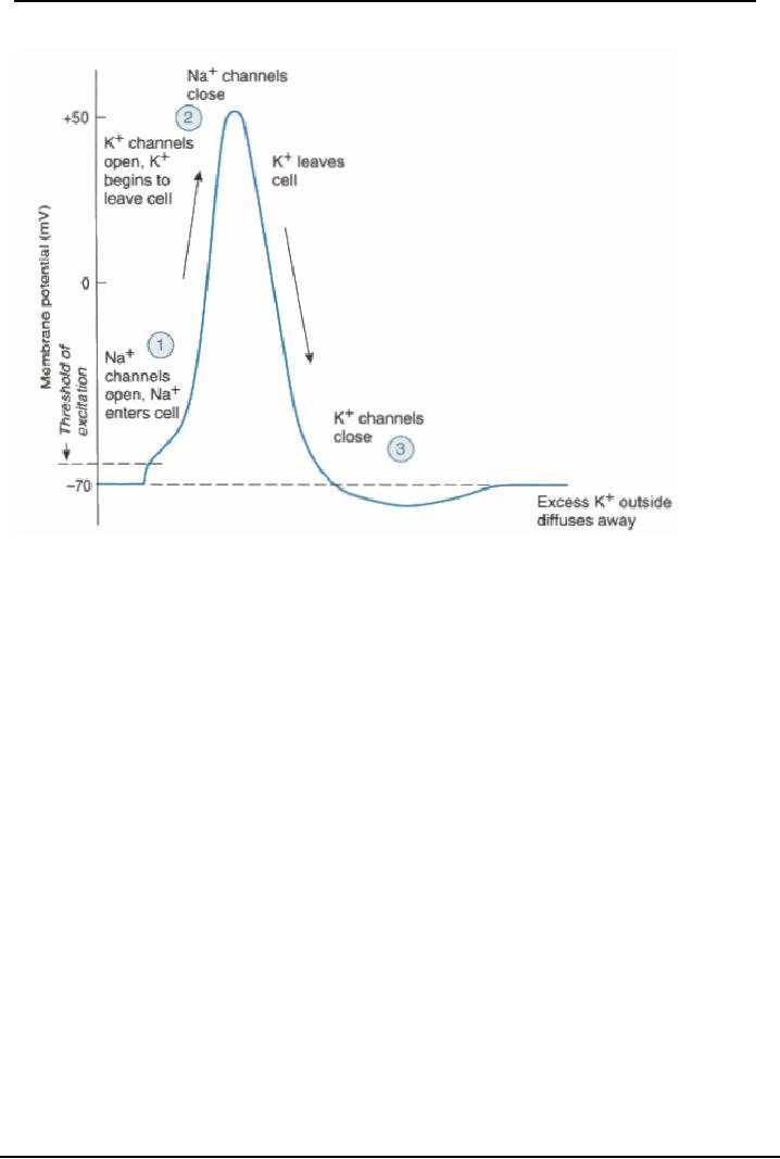
of charges in the household battery). This is known as the Resting Potential of
the neuron. At
this
stage the cell is at a Resting state. When positively charged ions enter the
cell, the inside becomes
positively
charged as compared to the outside, and the charge is recorded at +50 mV, the
cell will fire an
action
potential. The voltage difference is about 120 mV to get to an Action Potential
(How?).
ION
Concentration
Concentration Cell State
Inside
outside
+
Resting,
(impermeable to NA+ inside
the
50
460
Sodium
NA
(
large
cytoplasm
molecule)
Potassium
K+
400
10
Resting,
small molecule, moves in and out
(Small
molecule)
87

Neurological
Basis of Behavior (PSY -
610)
VU
Cloride
CL-
Resting,
small molecule moves in and out of the
40
560
cell
(Small
molecule)
Anions
A-
Resting
(impermeable to A- outside
the
345
0
(large
molecule)
cytoplasm)
From
Brown and Wallace (1980), and Carlson (1988), page 18
As
we can see there is a high
concentration of negatively charged
molecules inside the cell, and
these
ions
are trying to equalize the
two sides of the cellular
membrane.
Ionic
movement follows two processes to
maintain equilibrium and thereby causing
the movement of
electrical
charge. Ions move along
their osmotic/concentration gradient and
electrostatic gradient. When
molecules
move from areas of high
concentration to areas of low
concentration to create
equilibrium
especially
in a permeable or a semi permeable membrane
this process is known as
osmosis (nature
strives
for equilibrium). Therefore if
the concentration of ions is
low on one side the ions
would move to
equalize
the balance on both sides.
From the above table we can
see that all the four
would move to
equalize
concentrations. This is known as the
osmotic
gradient.
Similarly,
the law in electricity is that like charges repel and unlike charges attract,
therefore molecules
would
move towards balancing the electrostatic gradient.
Both
the forces of osmosis and electrostatic
gradient are working
together continuously to create
a
constant
state of movement of ions.
As
an example let's take a
glass of water, divide it
with a fine muslin cloth
(or sieve) drop a teaspoon
of
salt
(sodium chloride = NA+ CL-) on one side only.
There would be diffusion as the molecules
move to
equalize
both sides as one side has
both NA and CL and other does
not. Therefore both NA and
CL ions
would
move to equalize both sides
of the glass moving according to
their Osmotic gradient i.e.
to
equalize
and balance concentration.
However, the sieve does not
allow large ions to pass,
therefore large
ions
get stuck on side and the small ions
move to other side, leaving
CL- on one side and NA+ on the
other.
Now we see the electrostatic
gradient come
into action, as there are
negatively charged
molecules
on one side and the positively charged on
the other. This leads to
attraction and movement of
ions
again. However, only the smaller
positively charged molecules can
cross over. Thus, in
turn
osmotic
gradient moves ions to
equalize, then negatively
charged attract to move ions
again. This
movement
across the sieve causes flux in the
glass.
This
is the same kind of action
taking place in the neuronal/axonal
membrane leading to the resting
and
the
action potential.
In
the resting state of the axon the
membrane is impermeable to both
large ions a) positively
charged
sodium
ions (which are outside) and
the Anions (which are
inside) and the smaller chloride
(negative)
and
Potassium (positive) are
continuously moving back and forth
according to the osmotic and
electrostatic
gradients. However, this changes
when the axons receives
inputs from the cell soma to
fire,
there
is a change in the concentration of ions
as the cell membrane becomes
permeable and large
sodium
ions rush in, making in the
inside of the cell positively
charged.
Sodium
potassium pump:
When
the cell permeability changes,
large ions rush in NA+,
inside becomes positively
charged. The
cell
becomes impermeable again,
but it is stuck with the large
sodium ions inside. Then,
the cell
membrane
uses a biological pump known
as the sodium- potassium pump to
push out the NA+ and
carry
molecules of potassium back inside the
cell. This uses up to 40% of
the cell's energy as the cell
is
pushing
them against their osmotic gradients.
88
Neurological
Basis of Behavior (PSY -
610)
VU
How
does the resting potential
change to an action potential.
The cell at the resting
state is receiving
inputs
form all over which
are being summated at the
axonal hillock. There are
changes in the cell's
electrical
threshold that are taking
place.
The
inside is negative as compared to the
outside membrane, and the difference is
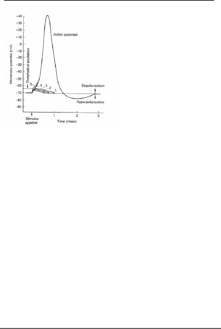
of -70 mV. This
negativity
can increase resulting in
Hyperpolarization is where there is an increase
negativity from -70
to
-80. On the other hand the Depolarization
are decreases in negativity
from -70 to -65, or -60 (
these
are
small depolarization) but a
larger depolarization of leads to
crossing the threshold and going
upto
+50mV.
This is an action potential
which leads the cell to
fire. Once the peak AP is reached, the
inside
electrical
charge starts becoming
negative, to the point that it
drops below the -70
mV.
After
action potential has been
fired, the cell goes into a
refractory state- hyperpolarized- to
about -75
mV.
It will not fire, till it
returns to the resting state
The
action potential lasts for
about 1/1000th of a second, and the refractory
period can continue for
about
some
milliseconds
89
Neurological
Basis of Behavior (PSY -
610)
VU
Firing
of the action potential leads to the
conductance of the signal. The rate and
speed of conductance
is
equivalent to 224 miles/hour
which is equal to 100meters per
sec in cat brain, in humans
it is about 60
meters
per second.
The
axonal conduction is an all-
or- none phenomenon, the cell would
fire an action potential
once the
threshold
is reached. The action would
be completed once it begins.
Once
the axonal transmission has crossed to
the postsynaptic site, it can lead to two
types of action:
The
Excitatory Post Synaptic
Potentials (EPSPs), this
would cause the post
synaptic site to fire an
action
potential.
This stimulates action in the post
synaptic site.
Inhibitory
Post Synaptic Potential (IPSPs)
inhibits ongoing firing of the
cell that it synapses on to.
So
activity
of the cell would be brought to a
resting state.
Since
there are multiple synapses on
each cell ( at the dendrites, the cell
soma), there may be
some
which
are IPSP and some which
are EPSP's, these
stimulations are summated
and if the stimulation
crosses
the excitatory threshold to arouse the
cell , it would fire
otherwise it would stay in the
resting
state.
Spatial
and temporal summation: multiple
synapses are continuously
adding together the EPSP's
and
IPSPs
received by them. There are
two kinds of summations of
stimulation that are carried
out at the
cell
soma and the axonal
hillock,
A)
Spatial summation: When a
neuron receives inputs from
several locations these can
EPSP's which
create
depolarization and IPSP's which
lead to hyper polarization.
These spread across the
cell
membrane
and reach the axonal hillock at the
same time they are
integrated and summated
algebraically
if
the sum is slightly negative
then a small hyperpolarization
would take place and the cell
would go
from
-70mV to -75 mV.
B)
Temporal Summation: When a
neuron receives input from
the same location but
repeatedly over
time
(could be EPSP's or IPSP's) the
are summed together received
one after another ( how can
this
happen
one stimulation is received and has
still not faded away, the
2nd one
received adds up as
does
90

Neurological
Basis of Behavior (PSY -
610)
VU
the
third and the fourth one).
After summation at the axonal hillock,
the neuron may either
depolarize
further
or hyperpolarize
Basic
Neuroanatomy: Anatomical Axis, Directions
and Planes of
Reference
Before
we study the brain we have to understand the
basic concepts of the locations,
sites and their
relationship
to each other is defined.
Just as we use the
directional reference of North-South, and
East-
West
in Geography, we also have specialized
terms for identifying the
directions in the brain
Basic
neuroanatomical axis: Anterior- posterior, dorsal- ventral, lateral
-medial;
In
humans we follow the same system that is followed for all other animals,
especially the vertebrates.
Anterior-posterior:
Anterior
towards the front: the nose end, and posterior is towards back,: the
tail
end,
so all structures in the front would be anteriorlly located and the structures
in the back would be
posteriorly
located. This is also known as the rostral-caudal axis (rostral: towards the
face and caudal:
towards
the tail, easier in animals which have tails!)
Dorsal-
Ventral: This axis is easier to
understand with a four legged animal or the fish than in
humans.
Dorsal
means towards the back for example the dorsal fin of shark of head and body,
ventral is towards
the
chest /stomach region or the bottom of the head. In humans the dorsal surface
becomes the back side
as
we stand. The top of the head, the back side facing the vertebral column are
then the dorsal areas
Medial-
Lateral: The third axis in which
medial is used as reference for areas towards the center or
the
mid
line. The nose is medially located with reference to the face and ear are
laterally located that is they
are
located toward the sides. Therefore the brain areas towards the outside are
laterally located. It is
important
to remember the other terms of reference which are continuously being used with
reference to
the
brain and various neuroanatomical sites
Ascending-
descending fibres: Descending refers to the
groups of nerves/ processes which travel down
from
the higher areas to lower areas: from cortex, the nerves descend to the Thalamus
and from the
thalamus
to the lower areas. Ascending refers to the nerves and the projections which
carry messages up
to
the higher brain areas.
Superior-inferior:
Superior
is those structures, nerve fibres or projections which lie on the top,
whereas
the
lower structures, projections, fibres, areas are referred to as inferior
(because they lie lower than, not
because
their functioning is lower).
Proximal-distal:
proximal
areas are those which lie closer to the brain or to each other. Those
areas
which
are farther are known as distally located areas. Ipsilateral-contralateral: Ipsi
means the same side
and
contra means the opposite side. Therefore ipsilateral would means those areas,
or fibres, or nerves
or
structures which are on the same side, whereas the contra lateral would be
structures, fibres or areas
on
the opposite side. The ipsilateral fibres would travel from the left side
occipital cortex to the left eye;
contralateral
would cross over at the optic chiasm to the right eye. Afferent- Efferent:
afferent are those
which
are bringing messages into the brain: these refer to the nerves which carry
information to the
brain
form the sensory areas Efferent taking info out of the brain or carry commands
messages from the
brain
to motor areas.
Planes
of reference: When brain is dissected
for studying the sections are cut and referred to in planes
of
reference. Horizontal sections are cut slicing the brain through from the dorsal
to the ventral areas.
(or
vice-versa) the sagittal cuts are made when we move in the lateral to the
medial- lateral direction.
The
mid sagittal section is made through the middle of the two hemispheres at the
level of the point of
joining.
The frontal section is cut from the front of the brain towards the
back.
91

Neurological
Basis of Behavior (PSY -
610)
VU
References:
1.
Kalat
J.W (1998) Biological Psychology
Brooks/ Cole
Publishing
2.
Carlson
N.R. (2005) Foundations of
Physiological Psychology Allyn and Bacon,
Boston
Pinel,
John P.J. (2003) Biopsychology
(5th edition) Allyn and Bacon
Singapore
3.
4.
Bloom
F, Nelson and Lazerson (2001), Behavioral
Neuroscience: Brain, Mind and Behaviors
(3rd
edition)
Worth Publishers New
York
5.
Bridgeman,
B (1988) The Biology of
Behaviour and Mind. John Wiley
and Sons New
York
6.
Brown,T.S.and
Wallace. (1980) P.M Physiological
Psychology0
Academic
Press New York
Note:
References
2, 3, 4, 7 more closely followed in
addition to the references cited in
text.
92
Table of Contents:
- INTRODUCTION:Descriptive, Experimental and/ or Natural Studies
- BRIEF HISTORICAL REVIEW:Roots of Behavioural Neurosciences
- SUB-SPECIALIZATIONS WITHIN THE BEHAVIORAL NEUROSCIENCES
- RESEARCH IN BEHAVIOURAL NEUROSCIENCES:Animal Subjects, Experimental Method
- EVOLUTIONARY AND GENETIC BASIS OF BEHAVIOUR:Species specific
- EVOLUTIONARY AND GENETIC BASIS OF BEHAVIOUR:Decent With Modification
- EVOLUTIONARY AND GENETIC BASIS OF BEHAVIOUR:Stereoscopic vision
- GENES AND EXPERIENCE:Fixed Pattern, Proteins, Genotype, Phenotypic
- GENES AND EXPERIENCE:Mendelian Genetics, DNA, Sex Influenced Traits
- GENES AND EXPERIENCE:Genetic Basis of behavior, In breeding
- GENES AND EXPERIENCE:Hybrid vigor, Chromosomal Abnormalities
- GENES AND EXPERIENCE:Behavioral Characteristics, Alcoholism
- RESEARCH METHODS AND TECHNIQUES OF ASSESSMENT OF BRAIN FUNCTION
- RESEARCH METHODS AND TECHNIQUES OF ASSESSMENT OF BRAIN FUNCTION:Activating brain
- RESEARCH METHODS AND TECHNIQUES OF ASSESSMENT OF BRAIN FUNCTION:Macro electrodes
- RESEARCH METHODS AND TECHNIQUES OF ASSESSMENT OF BRAIN FUNCTION:Water Mazes.
- DEVELOPMENT OF THE NERVOUS SYSTEM:Operation Head Start
- DEVELOPMENT OF THE NERVOUS SYSTEM:Teratology studies, Aristotle
- DEVELOPMENT OF THE NERVOUS SYSTEM:Stages of development, Neurulation
- DEVELOPMENT OF THE NERVOUS SYSTEM:Cell competition, Synaptic Rearrangement
- DEVELOPMENT OF THE NERVOUS SYSTEM:The issues still remain
- DEVELOPMENT OF THE NERVOUS SYSTEM:Post natal
- DEVELOPMENT OF THE NERVOUS SYSTEM:Oxygen level
- Basic Neuroanatomy:Brain and spinal cord, Glial cells, Oligodendrocytes
- Basic Neuroanatomy:Neuron Structure, Cell Soma, Cytoplasm, Nucleolus
- Basic Neuroanatomy:Control of molecules, Electrical charges, Proximal-distal
- Basic Neuroanatomy:Telencephalon, Mesencephalon. Myelencephalon
- Basic Neuroanatomy:Tegmentum, Substantia Nigra, MID BRAIN areas
- Basic Neuroanatomy:Diencephalon, Hypothalmus, Telencephalon, Frontal Lobe
- Basic Neurochemistry:Neurochemicals, Neuromodulator, Synaptic cleft
- Basic Neurochemistry:Changes in ionic gates, The direct method, Methods of Locating NT
- Basic Neurochemistry:Major Neurotransmitters, Mesolimbic, Metabolic degradation
- Basic Neurochemistry:Norepinephrine/ Noradrenaline, NA synthesis, Noadrenergic Pathways
- Basic Neurochemistry:NA and Feeding, NE and self stimulation: ICS
- Basic Neurochemistry:5HT and Behaviors, Serotonin and sleep, Other behaviours
- Basic Neurochemistry:ACH and Behaviors, Arousal, Drinking, Sham rage and attack
- Brain and Motivational States:Homeostasis, Temperature Regulation, Ectotherms
- Brain and Motivational States:Biological Rhythms, Circadian rhythms, Hunger/Feeding
- Brain and Motivational States:Gastric factors, Lipostatic theory, Neural Control of feeding
- Brain and Motivational States:Resting metabolic state, Individual differences
- Brain and Motivational States:Sleep and Dreams, Characteristics of sleep
- Higher Order Brain functions:Brain correlates, Language, Speech Comprehension
- Higher Order Brain functions:Aphasia and Dyslexia, Aphasias related to speech
- Higher Order Brain Functions:Principle of Mass Action, Long-term memory
- Higher Order Brain Functions:Brain correlates, Handedness, Frontal lobe