 |

Neurological
Basis of Behavior (PSY -
610)
VU
Lesson31
Basic
Neurochemistry
Objectives:
To
familiarize the students with
the
Various
NT and their role in the modulation of
behaviors
Classification
of Neurotransmitters. Monoamines:
Catechoalimnes
and
Indolemaine,
acetylcholine,
amino acid, and Peptide
Neurotransmitters
role in modulation of behaviors and
Aberration
Drugs
and Behavior:
Classification
of Psychopharmacological substances
Behavioral
correlates, Treatment:
Mechanism
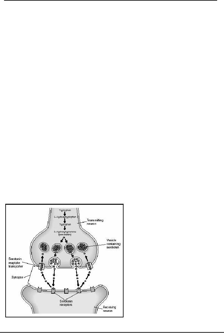
of synaptic transmission
Neurotransmitters:
synaptic transmission
Synaptic
transmission can be divided into several
clear cut and major steps these are
relatively
independent,
however each one step has to
occur before the next one
can take place
1.
Synthesis of the NT and
storage in the synaptic
vesicles. As we have
learnt earlier that
the
synaptic
vesicles are storage containers where NT
is protected from the deactivating
enzymes, thus
the
synaptic vesicles protect NT
from degradation by enzymes in cytoplasm.
These vesicles are
also
the
safe transporters of the NT. Where do they
come from? The synaptic
vesicles are manufactured
from
proteins in the cytoplasm of the cell
body by the Golgi apparatus.
These then travel
down
towards
the axonal endings to the synaptic buttons.
The packaging for Peptides
group of NTs
(which
are short chains of amino
acids) takes place in the Cisternas in
the synaptic buttons (button
or
bead like bulbous ends).
For the Non-Peptide class of
NTs, the packaging into
vesicles is carried
out
within the cytoplasm, before
they are transported down to the
axonal synaptic buttons.
The
material
can be transported in two different
directions by the axonal transport
system, the
anterograde
transport and the retrograde transport.
The
Anterograde
(forward)
axonal transport is a fast track transport
mechanism which
moves
materials out from the cell
body, through the microtubules towards
synaptic ending.
The
synaptic vesicles go through
this rapid system traveling
very fast speed of
400
millimiteres
per day. (It is like driving
in the fast lane). This is known as the
fast
anterograde
transport.
When
materials and synaptic vesicles ooze along the axon in the cytoplasm at a very
slow
speed
of less than 10 millimeters per day, they use the slow
anterograde transport
2.
Release of the Neurotransmitter: When the action potential
reaches the presynaptic ending it is
translated
into a chemical message (remember the neuronsa can communicate in both systems).
The
arrival
of the action potentials translated using the calcium gated channels. The
process is as
follows:
The action potential arrives at the terminal button; it leads to the opening of
the Calcium
channels.
This allows the calcium to get into the button and to trigger the release of
the
neurotransmitter.
If calcium is reduced in the extracellular space then the amount of NT is
also
reduced,
and it the extracellular Calcium is increased the amount of NT released is also
increased.
Release
is identified to occur through a process called exocytosis.
a)
The
NT vesicles move out towards the terminal to empty contents into the synaptic
cleft,
b)
The
NT vesicles fuse with presynaptic membrane, at contact the membrane opens up and
NT
molecules
are released into the cleft.
111

Neurological
Basis of Behavior (PSY -
610)
VU
c)
The
vesicle merges as part of the pre synaptic membrane, and the ruptureor the break
in the
membrane
eventually mends
Generation
of the post synaptic potential: this means action at the
receiving end at the post
3.
synaptic
potential after the NT molecule is received. When the NT molecule crosses over
the
synaptic
cleft (it is like crossing over a river full of alligators!) and gets
transferred to post
synaptic
membrane for action. There are several processes which would now take plce at
the post
receptor
sites
a)
Binding of NT molecules to post receptor site. All molecules released
would rush to reach
and
enter the postreceptor sites, the entry requires that they must connect or
"bind" chemically
with
the membrane site (a receptor protein). The membrane is very specialized with a
particular
configuration
therefore only those which resemble that shape and chemical composition
would
bind
these sites. This means the gates would open to allow only specific molecules to
enter
b)
Changes in ionic gates: The NT molecule leads to
changes in the chemically gated ionic
channels
in the receptor membrane for further action either through the direct or the
indirect
method,
i)
The direct method: The binding of the NT to
a receptor can directly open or close the
chemically
gated channels in the areas surrounding the membrane (to make it more permeable)
or
ii)
a
series of chemical changes can take place in the molecules in the cytoplasm
which can bring
about
a change in the status of the of the chemically gated ions channels of the
postreceptor site.
These
changes take place in chemicals/molecules (2nd messenger, Cyclic
Adenosine
MonoPhosphate
which is involved in conversion of Adenosine triphosphate to CAMP
through
enzymes.
Note:
the
Cycic AMP is needed for the energy in the cell for action). The second
method
uses
classof molecules which are called the 2nd messenger (because they are the ones which
intervenes
the messages and translate further action). The effect of CAMP is
brief...
Action
in the Post Receptor membrane: In the post synaptic
membrane, there are two kinds of
4
actions
that can take place, either an excitatory post synaptic potential
(EPSP)
or an Inhibitory
post
synaptic potential (IPSP').
The EPSP's generate an action potential in the post synaptic
membrane,and
the IPSP's inhibit ongoing activity in the cell membrane. Both of these
actions
depend
on a) the type of NT involved, i.e. some NT's are classified as excitatory NT's
and some
are
classified as inhibitory ( such as GABA), and , b) the site at which the action
is taking place.
The
NT action may be excitatory at some sites and inhibitory at other sites, as some
NT is
excitatory
at one site and inhibitory at another.
Inactivation
of the NT: What happens to an NT if
it is released from the vesicles
5.
a)
It
cannot stay in the neuron, or in the cleft,
b)
It
cannot keep activating the post synaptic membrane (otherwise one single dose of
amphetamine
stimulant
can last a life time!!!),
c)
It
cannot continue to stay in the cleft and keep the site full of molecules. This
would clutter the cleft
and
the sites. The NT has to be removed or degraded so that the systems remain
efficient and
clean.
There
are two well documented processes by which neurotransmitters are
deactivated:
·
Reuptake:
By
this mechanism the NT can return to the presynaptic areas and be taken in
for
recycling
and use. The reuptake processes allows the presynaptic area to reuptake and
absorb
the
molecules back. These are then repackaged into the vesicles and
used
·
Deactivating:
The
active chemical state or composition of the NT is deactivated by
chemicals/
enzymes
which are specialized to do this job. These enzymes locate free floating
unprotected
112

Neurological
Basis of Behavior (PSY -
610)
VU
NT
molecules in the synaptic cleft (and also in the presynaptic areas) to degrade
them and then
so
that they are excreted out of the cleft. Imagine that these are like the little
Pac men running
after
the little molecules.
6.
Recycling
of the vesicular membrane: The vesicles which had
ruptured are recycled. When many
synaptic
vesicles release molecules after fusing with the presynaptic membrane and the
process of
exocytosis,
the terminal button gets swollen with so many left over vesicles. Then the
pieces of excess
are
broken off and returned to cytoplasm. There these may be used again as packaging
material in one
of
the following forms;
a)
They
may be filled with
non-peptide NT by the cisternas.
b)
They
may be sent back to the cell
body by the retrograde transport (traveling at the
rate of 200
millimeters
per day.
c)
They
may be refilled with NT by the
golgi bodies in the cell
soma
d)
They
may be broken down and
molecules recycled.
Methods
of Locating NT:
Apart
from the many given techniques of
neuroanatomical tracing the following
techniques are
especially
used for the NT localization
identifying their sites and
their projections.
1.
Histoflouresence Technique: This was developed by
Falck and Hillarp around the early
1960's.
In this technique the monoamine group of NT's when exposed to formalin
fixative
glow
when exposed under a flourecent light. This technique was useful in locating the
various
monoamines,
their sites, their systems. However, this is none specialized as it does
not
differentiate
between various NT within the class of monoamines.
2.
.Receptor Binding
Autoradiography: The NT are radiolabelled
with a radio active isotope
(Hydrogen3
or carbon3) Then the neural tissue is exposed to the labeled ligand (molecule
that
binds
to a target). We can also inject this directly into the brain and expose the
slices for a
longer
period after decapitating the animal head and slicing the brain tissues. The
slices are
exposed
to a photographic plate which reacts to radioactivity and high radio active
areas show
up
in the plates.
3.
Monoclonal Antibodies: These involve
immunocytochemistry procedures. Just as the
lymphocytes
secrete antibodies, and hybrid lymphocytes and bone marrow cells
secrete
antibodies
and subdivide. We can use this same process to identify antibodies for
particular
proteins
(remember all NTs are chains of amino acids). Specific monoclonal antibodies
are
developed
and injected and they identify specific regions and target
proteins.
4.
Microiontophoresis (push pull cannulae). This procedure analyzes
the chemicals being
released
within the synapse. The response of the postsynaptic sites is monitored using a
double
barreled
pipette. The tip of the inner pipette (contains saline) is inserted into the
postsynaptic
membrane
to record intracellular voltage. Weak current when passed to stimulate the
neuronal
ending
leads to a discharge which is then pulled out for analysis, and also checked at
the
oscilloscope
for EPSP's or IPSP's
Major
Neurotransmitters:
There
are a large number of neurochemicals which have been classified as
neurotransmitters; there are
six
(6) major groups, and within each group there are several independent
neurotransmitters which have
specific
actions. These major groups are as follows:
113

Neurological
Basis of Behavior (PSY -
610)
VU
1.
Amino acids: These
are neurotransmitters which are
formed from chains of amino
acids, the
basis
of proteins. In this group the
major NT's are Glutamate,
Gamma aminobutyric
acid
(GABA),
Glycine (gly), and aspirate. This is the
largest group with relatively
quick acting
synaptic
connections. Glutamate is excitatory,
while GABA is known as the
inhibitory
neurotransmitter.
2.
Monoamines I: Catecholamines: This
group of neurotransmitters is synthesized from a
single
amino
acid; therefore this is
called mono (single) amine.
The monoamines modulate a
wide
range
of behaviors. The neurons of monoamines
have little bulbous bead-like
knobs throughout
the
length of the axons, through which the NT
appear to seep out. This
particular group of
Monoaminesis
called catecholamines because
they have one catechol group.
The
catecholamines
are Dopamine, Norepinephrine (also
known as Noradrenaline) and
Epinephrine
also
known as Adrenaline.
3.
Monoamines: Indoleamine: This
also belongs to the monoamine group, but
has a different
structure
attached to the amine group, the
indoleacetic acid. The
Indoleamine NT is known as
Serotonin
4.
Soluble gases: These
are small molecule NT. These
follow a different mechanism
of
transmission.
Since they are lipid soluble
they diffuse through the
cell membrane into
the
extracellular
space to pass into the other
cells. They work through
2nd messengers and break
down
immediately after action.
Nitric acid and Carbon
dioxide are two which have
been
discovered
so far.
5.
Acetylcholine: a small
molecule transmitter,one of its
kind--there are no other
NT's in this
group.
This is the only NT which
works on the neuromuscular joints
6.
Neuropeptides: a large
number of peptides (chain of 5 molecules) floating
around in the
brain-
and are possible candidates for NT
status. Among the well known
are the brain
opioids,
the
Endorphins ( large molecules) and
enkaphalins ( small molecules), the
pituitary peptides,
Substance
P and many others
www.chemistryexplained.com.NE-NA.Neurotranmitters
114

Neurological
Basis of Behavior (PSY -
610)
VU
References:
1.
Kalat J.W (1998) Biological
Psychology Brooks/ Cole
Publishing
2.
Carlson N.R. (2005) Foundations of
Physiological Psychology Allyn and Bacon,
Boston
3.
Pinel, John P.J. (2003)
Biopsychology (5th edition) Allyn and Bacon
Singapore
4
Bloom F, Nelson and Lazerson (2001),
Behavioral Neuroscience: Brain, Mind and
Behaviors (3rd
edition)
Worth Publishers New
York
5.
Bridgeman, B (1988) The
Biology of Behaviour and Mind. John
Wiley and Sons New
York
6.
Brown, T.S. and Wallace.(1980) P.M
Physiological Psychology
Academic
Press New York
7.
Seigel, G.J. (Ed. in chief)
Agranoff, B.W, Albers W.R.
and Molinoff, P.B. (Eds) (1989)
Basic
Neurochemistry:
Molecular, Cellular and Medical
Aspects
8.
Cooper, J.R, F.E Bloom,and
R.H Roth Biochemcial basis
of neuropharmacology 5th Edition,
OUP
Pharmacology,
Biochemcistray and behavior
(Additional
references for the module: Iversen and Iversen, Gazzaniga, Bloom, and
handouts)
Note:
References
2, 3, 4, 7 more closely followed in
addition to the references cited in
text.
115
Table of Contents:
- INTRODUCTION:Descriptive, Experimental and/ or Natural Studies
- BRIEF HISTORICAL REVIEW:Roots of Behavioural Neurosciences
- SUB-SPECIALIZATIONS WITHIN THE BEHAVIORAL NEUROSCIENCES
- RESEARCH IN BEHAVIOURAL NEUROSCIENCES:Animal Subjects, Experimental Method
- EVOLUTIONARY AND GENETIC BASIS OF BEHAVIOUR:Species specific
- EVOLUTIONARY AND GENETIC BASIS OF BEHAVIOUR:Decent With Modification
- EVOLUTIONARY AND GENETIC BASIS OF BEHAVIOUR:Stereoscopic vision
- GENES AND EXPERIENCE:Fixed Pattern, Proteins, Genotype, Phenotypic
- GENES AND EXPERIENCE:Mendelian Genetics, DNA, Sex Influenced Traits
- GENES AND EXPERIENCE:Genetic Basis of behavior, In breeding
- GENES AND EXPERIENCE:Hybrid vigor, Chromosomal Abnormalities
- GENES AND EXPERIENCE:Behavioral Characteristics, Alcoholism
- RESEARCH METHODS AND TECHNIQUES OF ASSESSMENT OF BRAIN FUNCTION
- RESEARCH METHODS AND TECHNIQUES OF ASSESSMENT OF BRAIN FUNCTION:Activating brain
- RESEARCH METHODS AND TECHNIQUES OF ASSESSMENT OF BRAIN FUNCTION:Macro electrodes
- RESEARCH METHODS AND TECHNIQUES OF ASSESSMENT OF BRAIN FUNCTION:Water Mazes.
- DEVELOPMENT OF THE NERVOUS SYSTEM:Operation Head Start
- DEVELOPMENT OF THE NERVOUS SYSTEM:Teratology studies, Aristotle
- DEVELOPMENT OF THE NERVOUS SYSTEM:Stages of development, Neurulation
- DEVELOPMENT OF THE NERVOUS SYSTEM:Cell competition, Synaptic Rearrangement
- DEVELOPMENT OF THE NERVOUS SYSTEM:The issues still remain
- DEVELOPMENT OF THE NERVOUS SYSTEM:Post natal
- DEVELOPMENT OF THE NERVOUS SYSTEM:Oxygen level
- Basic Neuroanatomy:Brain and spinal cord, Glial cells, Oligodendrocytes
- Basic Neuroanatomy:Neuron Structure, Cell Soma, Cytoplasm, Nucleolus
- Basic Neuroanatomy:Control of molecules, Electrical charges, Proximal-distal
- Basic Neuroanatomy:Telencephalon, Mesencephalon. Myelencephalon
- Basic Neuroanatomy:Tegmentum, Substantia Nigra, MID BRAIN areas
- Basic Neuroanatomy:Diencephalon, Hypothalmus, Telencephalon, Frontal Lobe
- Basic Neurochemistry:Neurochemicals, Neuromodulator, Synaptic cleft
- Basic Neurochemistry:Changes in ionic gates, The direct method, Methods of Locating NT
- Basic Neurochemistry:Major Neurotransmitters, Mesolimbic, Metabolic degradation
- Basic Neurochemistry:Norepinephrine/ Noradrenaline, NA synthesis, Noadrenergic Pathways
- Basic Neurochemistry:NA and Feeding, NE and self stimulation: ICS
- Basic Neurochemistry:5HT and Behaviors, Serotonin and sleep, Other behaviours
- Basic Neurochemistry:ACH and Behaviors, Arousal, Drinking, Sham rage and attack
- Brain and Motivational States:Homeostasis, Temperature Regulation, Ectotherms
- Brain and Motivational States:Biological Rhythms, Circadian rhythms, Hunger/Feeding
- Brain and Motivational States:Gastric factors, Lipostatic theory, Neural Control of feeding
- Brain and Motivational States:Resting metabolic state, Individual differences
- Brain and Motivational States:Sleep and Dreams, Characteristics of sleep
- Higher Order Brain functions:Brain correlates, Language, Speech Comprehension
- Higher Order Brain functions:Aphasia and Dyslexia, Aphasias related to speech
- Higher Order Brain Functions:Principle of Mass Action, Long-term memory
- Higher Order Brain Functions:Brain correlates, Handedness, Frontal lobe