 |

Introduction
to Psychology PSY101
VU
Lesson
13
NERVOUS
SYSTEM (2)
Membranes
of the Brain
�
Between
the surfaces of the brain and the skull,
there are three layers of
membrane called the
meanings,
which completely cover the brain
and spinal cord.
�
These
three membranes are:
1.
Dura
Matter
2.
Arachnoid
3.
Pia
Matter
Cerebrospinal
Fluid (CSF)
�
The
subarachnoid space contains a
fluid called cerebrospinal
fluid (CSF), a clear,
colorless
fluid covering the entire surface of
central nervous
system.
�
The
total volume of CSF is
125-150 ml.
�
Total
production of CSF is about
400-500 ml/day (about
0.36ml/min).
Association
Areas
�
Areas
in the cerebral cortex that are
not involved in primary motor
and sensory functions; rather
they
are involved in higher mental
functions such as learning, remembering,
thinking and
speaking.
�
Association
areas in the Frontal Lobes
are concerned with judging
and planning;
�
Damage
may lead to intact memory
but inability to plan out
something. Personality
�
may
also be affected.
�
Association
areas of other lobes are
related to other mental
functions; i.e. Temporal
Lobe enables us to
recognize
faces; damage to this area
causes inability to identify people
(although facial features
can be
described),
and gender and approximate
age too.
�
Association
areas in the posterior lobes are
involved in perception and memory. Damage
leads to
difficulty
in perceiving speech.
Spinal
Cord
�
Continuation
of the Medulla Oblongata.
�
The
spinal cord is about 45 cm long in men
and 43 cm long in women and
weighs about 35-40
grams.
�
The
vertebral column (back bone), encapsulating the
spinal cord, is about 70 cm
long
comprising
vertebra in the vertebral column.
�
The
spinal cord is much shorter than the vertebral
column.
�
Signals
arising in the motor areas of the
brain travel back down the cord
and leave in the
motor
neurons.
�
The
spinal cord also acts as a
minor coordinating center
responsible for some simple
reflexes
like
the withdrawal reflex.
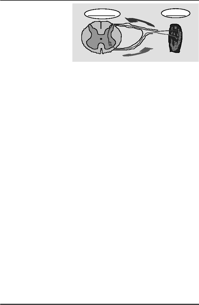
Reflex
- rapid
(and unconscious) response to
changes in the internal or external
environment, needed to maintain
homeostasis
Reflex
arc: the
neural pathway over which
impulses travel during a reflex. The
components of a reflex
arc
include:
1.
Receptor
-
responds to the stimulus
2.
Afferent
pathway --
sensory neuron
3.
Central
Nervous System
77

Introduction
to Psychology PSY101
VU
Peripheral
Nervous System (PNS)
Muscle
Spinal
Cord
Consists
of the spinal and
cranial
nerves;
these connect the CNS to
the
rest of the body. PNS
connects
the
body's sensory receptors to the
CNS,
and the CNS to the muscles
and
glands.
Parts
of Peripheral Nervous System
PNS
has two important
parts
1.
Skeletal/Somatic Nervous
System
�
Controls
the voluntary movements of our
skeletal muscles.
�
It reports
the current state of skeletal muscles
and carries instructions
back.
2.
Autonomic Nervous System
(ANS)
�
Considered
as the "self governing or self-regulatory
mechanism" because of its
involuntary
operation.
�
Controls
the glands and muscles of
internal organs e.g. heart,
stomach, and glandular
activity.
�
A.N.S.
has a dual function; i.e.
both arousing and
calming.
�
Comprises
two sub systems; Sympathetic
and parasympathetic nervous
systems.
a.
Sympathetic Nervous System
(SNS)
�
This
part of ANS arouses us for
defensive action.... fight or
flight.
�
If
something alarms, endangers,
excites, or enrages a person, the
sympathetic nervous
system
accelerates heart beat,
slows digestion, raises the sugar level
in blood, dilates the
arteries
and cools the body through
perspiration; makes one alert
and ready for action.
b.
Parasympathetic Nervous System
(PNS)
When
the stressful situation
subsides, parasympathetic nervous
system begins its
activity.
�
It
produces an effect opposite to that of
sympathetic nervous
system.
�
It
conserves energy by decreasing
heart beat, lowering blood
pressure, lowering blood
sugar
and
so on.
In
daily life situations, both
sympathetic and parasympathetic
systems work together to keep us in
steady
internal
state maintaining the homeostasis.
Studying
the Structure and Function of
the Brain
�
Electroencephalogram
(EEG): recording of the
electrical signals being transmitted
within
the
brain, through electrodes attached to the
skull.
�
Computerized
Axial Tomography (CAT): a computer
constructs an image of the brain
by
combining
thousands of separate X-rays taken
from slightly different
angles.
�
Magnetic
Resonance Imaging (MRI): the
scan produces a powerful
magnetic field to
provide
a computer generated, detailed image of
the structure of the brain.
�
Super
Conducting Quantum Interference Device
(SQUID): a scan
sensitive to minute
changes
in the magnetic field occurring when
neurons are firing.
�
Positron
Emission Tomography
(PET):
a scan
showing biochemical
activity
within
the brain at any given
moment.
78
Table of Contents:
- WHAT IS PSYCHOLOGY?:Theoretical perspectives of psychology
- HISTORICAL ROOTS OF MODERN PSYCHOLOGY:HIPPOCRATES, PLATO
- SCHOOLS OF THOUGHT:Biological Approach, Psychodynamic Approach
- PERSPECTIVE/MODEL/APPROACH:Narcosis, Chemotherapy
- THE PSYCHODYNAMIC APPROACH/ MODEL:Psychic Determinism, Preconscious
- BEHAVIORAL APPROACH:Behaviorist Analysis, Basic Terminology, Basic Terminology
- THE HUMANISTIC APPROACH AND THE COGNITIVE APPROACH:Rogers’ Approach
- RESEARCH METHODS IN PSYCHOLOGY (I):Scientific Nature of Psychology
- RESEARCH METHODS IN PSYCHOLOGY (II):Experimental Research
- PHYSICAL DEVELOPMENT AND NATURE NURTURE ISSUE:Nature versus Nurture
- COGNITIVE DEVELOPMENT:Socio- Cultural Factor, The Individual and the Group
- NERVOUS SYSTEM (1):Biological Bases of Behavior, Terminal Buttons
- NERVOUS SYSTEM (2):Membranes of the Brain, Association Areas, Spinal Cord
- ENDOCRINE SYSTEM:Pineal Gland, Pituitary Gland, Dwarfism
- SENSATION:The Human Eye, Cornea, Sclera, Pupil, Iris, Lens
- HEARING (AUDITION) AND BALANCE:The Outer Ear, Auditory Canal
- PERCEPTION I:Max Wertheimer, Figure and Ground, Law of Closure
- PERCEPTION II:Depth Perception, Relative Height, Linear Perspective
- ALTERED STATES OF CONSCIOUSNESS:Electroencephalogram, Hypnosis
- LEARNING:Motor Learning, Problem Solving, Basic Terminology, Conditioning
- OPERANT CONDITIONING:Negative Rein forcer, Punishment, No reinforcement
- COGNITIVE APPROACH:Approach to Learning, Observational Learning
- MEMORY I:Functions of Memory, Encoding and Recoding, Retrieval
- MEMORY II:Long-Term Memory, Declarative Memory, Procedural Memory
- MEMORY III:Memory Disorders/Dysfunctions, Amnesia, Dementia
- SECONDARY/ LEARNT/ PSYCHOLOGICAL MOTIVES:Curiosity, Need for affiliation
- EMOTIONS I:Defining Emotions, Behavioral component, Cognitive component
- EMOTIONS II:Respiratory Changes, Pupillometrics, Glandular Responses
- COGNITION AND THINKING:Cognitive Psychology, Mental Images, Concepts
- THINKING, REASONING, PROBLEM- SOLVING AND CREATIVITY:Mental shortcuts
- PERSONALITY I:Definition of Personality, Theories of Personality
- PERSONALITY II:Surface traits, Source Traits, For learning theorists, Albert Bandura
- PERSONALITY III:Assessment of Personality, Interview, Behavioral Assessment
- INTELLIGENCE:The History of Measurement of Intelligence, Later Revisions
- PSYCHOPATHOLOGY:Plato, Aristotle, Asclepiades, In The Middle Ages
- ABNORMAL BEHAVIOR I:Medical Perspective, Psychodynamic Perspective
- ABNORMAL BEHAVIOR II:Hypochondriasis, Conversion Disorders, Causes include
- PSYCHOTHERAPY I:Psychotherapeutic Orientations, Clinical Psychologists
- PSYCHOTHERAPY II:Behavior Modification, Shaping, Humanistic Therapies
- POPULAR AREAS OF PSYCHOLOGY:ABC MODEL, Factors affecting attitude change
- HEALTH PSYCHOLOGY:Understanding Health, Observational Learning
- INDUSTRIAL/ORGANIZATIONAL PSYCHOLOGY:‘Hard’ Criteria and ‘Soft’ Criteria
- CONSUMER PSYCHOLOGY:Focus of Interest, Consumer Psychologist
- SPORT PSYCHOLOGY:Some Research Findings, Arousal level
- FORENSIC PSYCHOLOGY:Origin and History of Forensic Psychology