 |

Health
Psychology PSY408
VU
LESSON
07
THE
FUNCTION OF NERVOUS
SYSTEM
Prologue
When
Tom was born 20 years
ago, his parents were
thrilled. Here was their
first child-- a delightful
baby
with
such promise for the future.
He seemed to be healthy. His parents
were pleased that he began
to
consume
large amounts of milk, often
without becoming satiated. They
took this as a good sign.
But, in this
case,
it wasn't.
As
the weeks went by, Tom's
parents noticed that he wasn't gaining as
much weight as he should,
especially
since
he was still consuming lots of
milk. He started to cough
and wheeze often and
developed one
respiratory
infection after another. They
became concerned, and so did
his pediatrician. After a series
of
tests,
the devastating diagnosis was
clear: Tom had Cystic
Fibrosis, a chronic,
progressive, and eventually fatal
disease.
Cystic fibrosis is an inherited disease
of the respiratory system for which
there is no cure and
no
effective
treatment.
Tom
has had a difficult life,
and so has his family.
The respiratory infections he had in infancy
were just the
beginning.
His disease causes thick,
sticky secretions that constantly
block airways, trap air in the
lungs, and
help
bacteria to thrive. Other
body systems also become
affected, causing additional
problems, such as
insufficient
absorption of food and vitamins. As a
result, he was sick often
and remained short,
underweight,
and weak compared with
other children. His social relationships
have always been limited
and
strained,
and the burden of his illness
has taken its toll on
his parents.
When
Tom was younger and people
asked him, "what do you want
to be when you grow up'? he
would
answer,
I'm going to be an angel when I grow
up." What other plans could
he have had, realistically? At
20,
he
has reached the age by which
half of the victims of cystic fibrosis
die. Physical complications,
such as
heart
damage, that generally
afflict several body systems
in the last stages of this disease
have begun to
appear.
We
can see in Tom's story that
biological factors, such as
heredity, can affect health; illness
can alter social
relationships;
and all interrelated physiological
systems of the body can be
affected.
An
understanding of health requires that we
have a working knowledge of human
physiology. This
knowledge
makes it possible to understand,
among others, such issues as
how good health habits
make
illness
less likely, how genetic or
environmental factors influence the
physical body of human
beings etc.
In
this lecture we will talk about the
nervous system. In our coming
lectures we will outline the
other major
physical
systems of the body. Our
discussions will focus on the
normal functions of these
systems, but we
will
consider some important
problems, too. For example,
what determines the degree of paralysis a
person
suffers
after injury to the spine? How
does stress affect our body
systems? What is a heart
attack, and what
causes
it?
THE
NERVOUS SYSTEM
How
the Nervous System
Works
We
all know that the nervous
system, particularly the brain, in human
beings and other animals
controls the
way
we initiate behavior and
respond to events in our
world. The nervous system
receives information
about
changes in the environment from
sensory organs, including the
eyes, ears, and nose,
and it transmits
directions
that tell our muscles
and other internal organs
how to react. The brain
also stores
information--
being
a repository for our memory of
past events--and provides our capability
for thinking, reasoning,
and
creating.
28

Health
Psychology PSY408
VU
The
nervous system is constantly integrating the
actions of our internal
organs--although we are
not
generally
aware of it. Many of these
organs, such as the heart
and digestive tract, are
made of muscle
tissues
that
respond to commands. The
nervous system provides these
commands through an intricate network
of
billions
of specialized nerve cells,
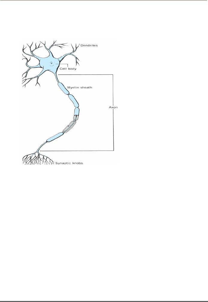
called neurons.
Although
neurons in different parts of the
nervous system have a variety of
shapes and sizes, the
diagram
on
your screen shows their
general features. Projecting from the
cell
body are
clusters of branches
called
dendrites.
Generally,
dendrites function as receivers
for messages from adjacent
neurons. These
messages
then
travel through a long, slender
projection called the axon,
which
splits into branches at the
Far end. The
tips
of these branches have small
swellings called synaptic
knobs that
connect to the dendrites of
other
neurons,
usually through a Fluid-filled
gap. This junction is called
a synapse.
Messages
From the knobs cross the gap to
adjacent neurons and in this
way eventually reach
their
destination.
What
changes occur in the nervous
system as a person develops? By the time
the typical baby is born, a
basic
structure has been formed
for almost all the neurons
this person will have. But
the nervous system is
still
quite immature--For instance, the
brain weighs only about 25%
of the weight it will have when
the
child
reaches adulthood. Most of the
growth in brain size after
birth results from an
increase in the number
of
Glial
cells and
the presence of a white fatty
substance called myelin.
The
Glial cells are thought to
service
and
maintain the neurons.
A
myelin sheath surrounds the axons of
most, but not all, neurons.
This sheath is responsible
for increasing
the
speed of nerve impulses and
preventing them from being interfered
with by adjacent nerve
impulses,
much
the way insulation is used on
electrical wiring. The importance of
myelin can be seen in the
disease
called
multiple
sclerosis,
which results when the myelin
sheath degenerates and
nerves become severed
(Trapp,
1998). People afflicted with
this disease have weak
muscles that lack
coordination and move
29

Health
Psychology PSY408
VU
spastically
(AMA, 1989).
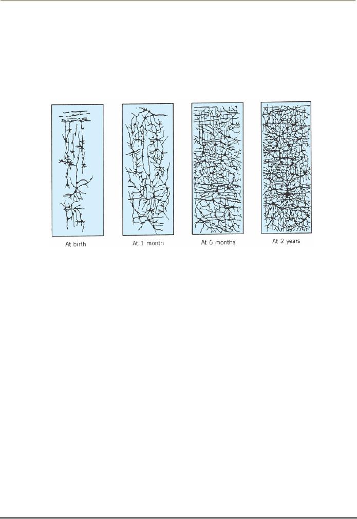
As
the infant grows, the network of
dendrites and synaptic knobs to
carry messages to and from
other
neurons
expands dramatically, as you can see in
the diagram.
The
myelin sheath covering the neurons is better developed
initially in the upper regions of the
body than in
the
lower regions. During the
first years of life, the
progress in myelin growth spreads
down the body--
from
the head to shoulders, to the arms
and hands, to the upper chest
and abdomen, and then the
legs and
feet.
This sequence is reflected in the
individual's motor development: the upper
parts of the body are
brought
under control at earlier ages
than the lower parts.
Studies with animals have
found that chronic
poor
nutrition early in life
impairs brain growth by retarding the
development of myelin, Glial cells,
and
dendrites.
Such impairment can produce long-lasting
deficits in a child's motor and intellectual.
Although
researchers
had thought that the brain
forms few, if any, new
neurons after birth, it is now
known that new
cells
do form in some areas of the brain,
but it is not yet clear
how extensive this growth
is.
Beginning
in early adulthood, the brain slowly
loses weight with age.
Although the number of brain
cells
does
not change very much, the
synapses do, leading to a
decline in ability to send
nerve impulses. These
alterations
in the brain are associated
with the declines people often notice in
their mental and
physical
functions
after they reach 50 or 60 years of
age.
The
nervous system is enormously complex
and basically has two major
divisions--the central
nervous
system
and the peripheral nervous system--that
connect to each other. The
central nervous system
consists
of
the brain and spinal cord.
The peripheral
nervous system
is composed of the remaining network of
neurons
throughout
the body. Each of these major divisions
consists of interconnected lower-order divisions
or
structures.
We will examine the nervous
system, beginning at the top and
working our way
down.
30

Health
Psychology PSY408
VU
The
Central Nervous
System
People's
brain and spinal cord race
toward maturity early in
life. For example, the brain
weighs 75% of its
adult
weight at about 2 years of age, 90% at 5
years, and 95% at 10 years
(Tanner, 1970, 1978). The
brain
may
be divided into three parts:
the forebrain, the
cerebellum,
and
the brainstem.
Each
of these parts has
special
functions.
The
Forebrain
The
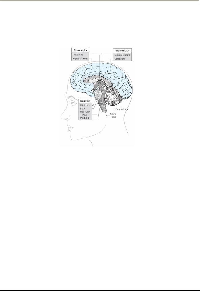
forebrain is the uppermost part of the
brain. As you can see the diagram on the
screen, the forebrain
has
two main subdivisions: the
telencephalon,
which
consists of the cerebrum and the
limbic system, and
the
diencephalon,
which
includes the thalamus and
hypothalamus. As a general rule, areas
toward the top and
outer
regions of the brain are
involved in our perceptual,
motor, learning, and conceptual
activities. Regions
toward
the center and bottom of the
brain are involved mainly in
controlling internal and
automatic body
functions
and
in
transmitting
information
to
and
from
the
telencephalon.
The
cerebrum is the upper and largest
portion of the human brain
and includes the cerebral
cortex, its
outermost
layer. The cerebrum controls complex
motor and mental activity.
It develops rapidly in the
first
few
years of life, becoming
larger, thicker, and more
convoluted. The cerebrum has
two halves--the left
hemisphere
and
the right
hemisphere--each of
which looks like the left
hemispheres shown
here.
31

Health
Psychology PSY408
VU
Although
the left and right
hemispheres are physically
alike, they control different
types of processes.
For
one
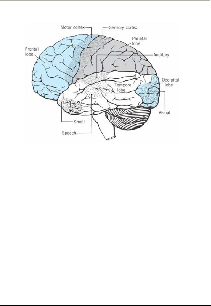
thing, the motor cortex of each
hemisphere controls motor movements on
the opposite side of the
body.
(You can see the next
diagram of brain showing the motor cortex
on your screen now). This is
why
damage
to the motor cortex on, say, the
right side of the brain may
leave part of the left side
of the body
paralyzed.
The two hemispheres also
control different aspects of
cognitive and language
processes. In most
people,
the left hemisphere contains the
areas that handle language
processes, including speech
and writing.
The
right hemisphere usually
processes such things as visual
imagery, emotions, and the perception
of
patterns,
such as melodies (Tortora &
Grabowski, 2000).
You
probably noticed in our previous diagram
that each hemisphere is
divided into a front part,
called the
frontal
lobe, and three back parts:
the temporal, occipital, and parietal lobes.
The frontal
lobe is
involved in a
variety
of functions, one of which is motor
activity. The back part of
the frontal lobe contains the
motor
cortex,
which controls the skeletal muscles of
the body. If a patient, who is undergoing
brain surgery,
receives
stimulation to the motor cortex, some
part of the body will move.
The frontal lobe is also
involved
in
important mental activities,
such as the association of ideas,
planning, self-awareness, and
emotion. As a
result,
injury to areas of this lobe
can produce personality and emotional
reactions.
The
temporal
lobe is
chiefly involved in hearing,
but also in vision and
memory. Damage to this region can
impair
the person's comprehension of speech
and ability to determine the
direction from which a sound
is
coming.
The
occipital
lobe contains
the principal visual area of the brain.
Damage to the occipital lobe can
produce
blindness
or the inability to recognize an object by
sight.
The
parietal
lobe is
involved mainly in body sensations,
such as of pain, cold, heat, touch, and
body
movement.
32

Health
Psychology PSY408
VU
The
second part of the telencephalon--called
the limbic system--lies along the innermost edge of
the
cerebrum,
and adjacent to the diencephalon (refer
back to diagram 3). The
limbic system is not
well
understood
yet. It consists of several
structures that seem to be
important in the expression of
emotions,
such
as fear, anger, and excitement. To the
extent that heredity affects a person's
emotions, it may do so by
determining
the structure arid function of the limbic
system.
The
diencephalon includes two structures--the
thalamus and hypothalamus--that
lie below and
are
partially
encircled by the limbic system.
The thalamus is a truly
pivotal structure in the flow of
information
in
the nervous system. It functions as the
chief relay station for directing sensory
messages, such as of pain
or
visual images, to appropriate points in
the cerebrum, such as the occipital or parietal lobe.
The thalamus
also
relays commands going out to the
skeletal muscles from the
motor cortex of the cerebrum.
The
hypothalamus, a small structure
just below the thalamus,
plays an important role in
people's emotions
and
motivation.
Its
function affects eating,
drinking, and sexual
activity, for instance
(Tortora & Grabowski, 2000).
For
example,
when the body lacks water or nutrients,
the hypothalamus detects this and
arouses the sensation of
thirst
or hunger, which is relieved when we
consume water or food.
Research with animals has
shown that
stimulation
of specific areas of this structure
can cause them to eat when
they are full and stop
eating when
they
are hungry. A rare disease
that affects this structure
can cause people to become overweight.
Another
important
function of the hypothalamus is to maintain
homeostasis--a
state
of balance or normal function
among
our body systems.
Our
normal body temperature and
heart rate, which are
characteristic of healthy individuals,
are examples of
homeostasis.
When our bodies are cold,
for instance, we shiver, thus
producing heat. When we are
very
warm,
we perspire, thus cooling the
body. The hypothalamus controls
these adjustments. We will see
later
that
the hypothalamus also plays an
important role in our reaction to
stress.
The
Cerebellum
The
cerebellum lies at the back of the brain,
below the cerebrum. The main
function of the cerebellum is in
maintaining
body balance and
coordinating movement. This structure
has nerve connections to the
motor
cortex
of the cerebrum and most
sense organs of the body.
When areas of the cerebrum
initiate specific
movements,
the cerebellum makes our
actions precise and well
coordinated.
How
does the cerebellum do this? There
are at least two ways.
First, it continuously compares our
intent
with
our performance, ensuring
that a movement goes in the right
direction, at the proper rate,
and with
appropriate
force. Second, it smoothes our
movements. Because of the forces
involved in movement,
there
is
an underlying tendency for
our motions to go quickly back
and forth, like a tremor.
The cerebellum
damps
this tendency. When injury
occurs to the cerebellum, the person's
actions become jerky
and
uncoordinated--a
condition called ataxia.
Simple
movements, such as walking or
touching an object,
become
difficult and
unsteady.
Diagram
3 (above) shows the location of the
cerebellum relative to the brainstem,
which is the next
section
of
the brain we will
discuss.
The
Brainstem
The
lowest portion of the brain--called the
brainstem--has the form of an oddly
shaped knob at the top
of
the
spinal cord. The brainstem
consists of four parts
midbrain, pons, reticular system,
and medulla.
The
midbrain lies at the top of the
brainstem. It connects directly to the
thalamus above it, which
relays
messages
to various parts of the forebrain.
The midbrain receives
information from the visual
and auditory
systems
and is especially important in
muscle movement. The disorder
called Parkinson's disease
results
from
degeneration
of an area of the midbrain (Tortora &
Grabowski, 2000). People severely
afflicted with this
33

Health
Psychology PSY408
VU
disease
have noticeable motor
tremors, and their neck
and trunk postures become
rigid, so that they walk in
a
crouch. Sometimes the tremors
are so continuous and vigorous that the
victim becomes crippled.
The
reticular system is a network of neurons
that extends from the bottom
to the top of the brainstem
and
into
the thalamus. The reticular system
plays an important role in
controlling our states of
sleep, arousal,
and
attention. When people suffer a coma,
often it is this system that is
injured or disordered. Epilepsy
a
condition
in which a victim may become
unconscious and begin to convulse
seems to involve an
abnormality
in the reticular system. One type of epileptic seizure
called grand
mal may
result from
reverberating
cycles" in the reticular system:
That
is, one portion of the
system stimulates another portion,
which stimulates a third
portion, and this in
turn
re-stimulates the first portion,
causing a cycle that
continues for 2 to 3 minutes,
until the neurons of
the
system fatigue so greatly that the
reverberation ceases. Following a grand
mal seizure, the person
often
sleeps
at least a few minutes and
sometimes for hours.
The
pons forms a large bulge at the
front of the brainstem and is
involved in eye movements,
facial
expressions,
and chewing. At the bottom of the
brain- stem is the medulla,
which contains vital centers
that
control
breathing, heartbeat rate, and the
diameter of blood vessels (which
affects blood pressure).
Because
of
the many vital functions it controls,
damage to the medulla can be
life threatening. Polio,
a
crippling
disease
that was once epidemic,
sometimes damaged the center
that controls breathing. Patients
suffering
such
damage needed constant
artificial respiration to breathe
(McClintic, 1985).
The
Spinal Cord
Extending
down the spine from the
brainstem is the spinal cord, a major
neural pathway that
transmits
messages
between the brain and
various parts of the body. It
contains neurons that carry
impulses away
from
(the efferent
direction)
and toward (afferent)
the brain.
Efferent commands travel down the cord on
their
way
to produce muscle action; afferent
impulses come to the spinal cord
from sense organs in all
parts of
the
body.
The
organization of the spinal cord parallels
that of the body--that is, the higher the
region of the cord, the
higher
the parts of the body to which it
connects. Damage to the spinal cord
results in impaired motor
function
or paralysis: the duration and extent of
the impairment depends on the amount and
location of
damage.
If the damage does not sever
the cord, the impairment is less severe,
and may be temporary. If the
lower
portion of the cord is severed, the lower
areas of the body are
paralyzed--a condition called
paraplegia.
If
the upper portion of the spinal cord is
severed, paralysis is more
extensive. Paralysis of the legs
and arms
is
called quadriplegia.
34
Table of Contents:
- INTRODUCTION TO HEALTH PSYCHOLOGY:Health and Wellness Defined
- INTRODUCTION TO HEALTH PSYCHOLOGY:Early Cultures, The Middle Ages
- INTRODUCTION TO HEALTH PSYCHOLOGY:Psychosomatic Medicine
- INTRODUCTION TO HEALTH PSYCHOLOGY:The Background to Biomedical Model
- INTRODUCTION TO HEALTH PSYCHOLOGY:THE LIFE-SPAN PERSPECTIVE
- HEALTH RELATED CAREERS:Nurses and Physician Assistants, Physical Therapists
- THE FUNCTION OF NERVOUS SYSTEM:Prologue, The Central Nervous System
- THE FUNCTION OF NERVOUS SYSTEM AND ENDOCRINE GLANDS:Other Glands
- DIGESTIVE AND RENAL SYSTEMS:THE DIGESTIVE SYSTEM, Digesting Food
- THE RESPIRATORY SYSTEM:The Heart and Blood Vessels, Blood Pressure
- BLOOD COMPOSITION:Formed Elements, Plasma, THE IMMUNE SYSTEM
- SOLDIERS OF THE IMMUNE SYSTEM:Less-Than-Optimal Defenses
- THE PHENOMENON OF STRESS:Experiencing Stress in our Lives, Primary Appraisal
- FACTORS THAT LEAD TO STRESSFUL APPRAISALS:Dimensions of Stress
- PSYCHOSOCIAL ASPECTS OF STRESS:Cognition and Stress, Emotions and Stress
- SOURCES OF STRESS:Sources in the Family, An Addition to the Family
- MEASURING STRESS:Environmental Stress, Physiological Arousal
- PSYCHOSOCIAL FACTORS THAT CAN MODIFY THE IMPACT OF STRESS ON HEALTH
- HOW STRESS AFFECTS HEALTH:Stress, Behavior and Illness, Psychoneuroimmunology
- COPING WITH STRESS:Prologue, Functions of Coping, Distancing
- REDUCING THE POTENTIAL FOR STRESS:Enhancing Social Support
- STRESS MANAGEMENT:Medication, Behavioral and Cognitive Methods
- THE PHENOMENON OF PAIN ITS NATURE AND TYPES:Perceiving Pain
- THE PHYSIOLOGY OF PAIN PERCEPTION:Phantom Limb Pain, Learning and Pain
- ASSESSING PAIN:Self-Report Methods, Behavioral Assessment Approaches
- DEALING WITH PAIN:Acute Clinical Pain, Chronic Clinical Pain
- ADJUSTING TO CHRONIC ILLNESSES:Shock, Encounter, Retreat
- THE COPING PROCESS IN PATIENTS OF CHRONIC ILLNESS:Asthma
- IMPACT OF DIFFERENT CHRONIC CONDITIONS:Psychosocial Factors in Epilepsy